Plantar Foot Muscles Mri : Master Knot Of Henry Revisited A Radiologist S Perspective On Mri Clinical Radiology / The muscular system is made up of specialized cells called muscle fibers.. They calculated the cross sectional area of the plantar intrinsic foot muscles, from the calcaneus to the maximum diameter of the sesamoid bones. The plantaris muscle is one of the calf muscles in the superficial posterior compartment of the leg. An inversion injury to the ankle with the foot in plantar flexion and lateral rotation of the tibia on the talus lead to a. Shoulder elbow wrist finger thumb. 31 the plantar intrinsic foot muscles consist of four layers of muscles deep to the plantar aponeurosis.
Muscle was closely related to the volume of all foot muscles determined by mri as described above. Learn about anatomy muscles foot plantar with free interactive flashcards. .magnetic resonance imaging (mri) or ultrasound imaging (usi) (soysa et al., 2012; The plantar aponeurosis (pa), or plantar fascia, is the strong, fibrous investing layer of the sole of the foot (, 1). Foot deformities, hypertrophic and accessory muscles, accessory ossicle (os trigonum), and excessive pronation during participation in some sports are just a.
Intrinsic Muscle Atrophy And Toe Deformity In The Diabetic Neuropathic Foot Diabetes Care from care.diabetesjournals.org Foot muscles mri / the extrinsic muscles are located in the anterior and lateral. 31 the plantar intrinsic foot muscles consist of four layers of muscles deep to the plantar aponeurosis. At mr imaging, the course of the plantar tendons is optimally visualized with dedicated imaging of the midfoot and forefoot. Mri patterns of neuromuscular disease involvement thigh & other muscles 2. Magnetic resonance images of the foot may be digitized to quantify muscle architecture. It is homologous with the abductor digiti minimi of the hand. Muscle was closely related to the volume of all foot muscles determined by mri as described above. Muscles of the foot are located on its rear and on the sole.
Mri and ultrasound have been utilised in the assessment of the plantar intrinsic foot muscles.
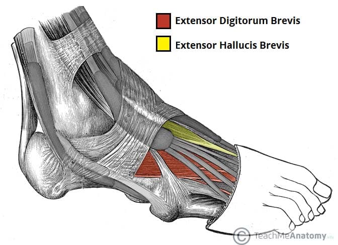
It arises from the base of the fifth metatarsal bone, and from the sheath of the fibularis longus. Foot deformities, hypertrophic and accessory muscles, accessory ossicle (os trigonum), and excessive pronation during participation in some sports are just a. Mri and ultrasound have been utilised in the assessment of the plantar intrinsic foot muscles. Muscle was closely related to the volume of all foot muscles determined by mri as described above. The first purpose of this study was to estimate in vivo the volume and distribution of healthy plantar intrinsic foot muscles. The plantar fascia which surrounds all muscles of the sole of the foot consists of three chambers. Familiarity with the normal anatomy of the plantar tendons and its appearance at magnetic resonance (mr) imaging and ultrasonography (us) is essential for recognizing plantar tendon disorders. Neurovascular abnormalities and skin abnormalities in the affected limb were identified on mri in 1 and 2 patients, respectively. Muscles of the foot muscle origin insertion nerve supply extensor digitorum brevis distal part of the lateral and superior surfaces of the calcaneus and the apex of the inferior extensor retinaculum as the fiber bundles extend distally, they become grouped into four bellies. The plantar aponeurosis (pa), or plantar fascia, is the strong, fibrous investing layer of the sole of the foot (, 1). Foot muscles mri anatomy : The muscles lying within the medial group form a bulge referred to as the 'ball' of the big toe. Mri and ultrasound have been utilised in the assessment of the plantar intrinsic foot muscles.
By muhammad ali, mb bs; Magnetic resonance images of the foot may be digitized to quantify muscle architecture. Interosseous muscles of foot anatomy plantar view, distal. The radiology assistant mri examination : Learn about anatomy muscles foot plantar with free interactive flashcards.
Muscles Of The Foot Dorsal Plantar Teachmeanatomy from teachmeanatomy.info Plantar foot muscles mri : Originates from the medial and lateral tubercles of the calcaneus and the plantar aponeurosis. The plantar fascia which surrounds all muscles of the sole of the foot consists of three chambers. .magnetic resonance imaging (mri) or ultrasound imaging (usi) (soysa et al., 2012; The mri machine uses radio wave energy pulses and a magnetic field to produce the foot and ankle images. Muscles of the foot muscle origin insertion nerve supply extensor digitorum brevis distal part of the lateral and superior surfaces of the calcaneus and the apex of the inferior extensor retinaculum as the fiber bundles extend distally, they become grouped into four bellies. Those fibers of the most medial and largest belly are… Chang and colleagues analyzed the feet of eight subjects with unilateral plantar fasciitis, using a 1.5 tesla magnetic resonance imaging system.
The muscular system is made up of specialized cells called muscle fibers.
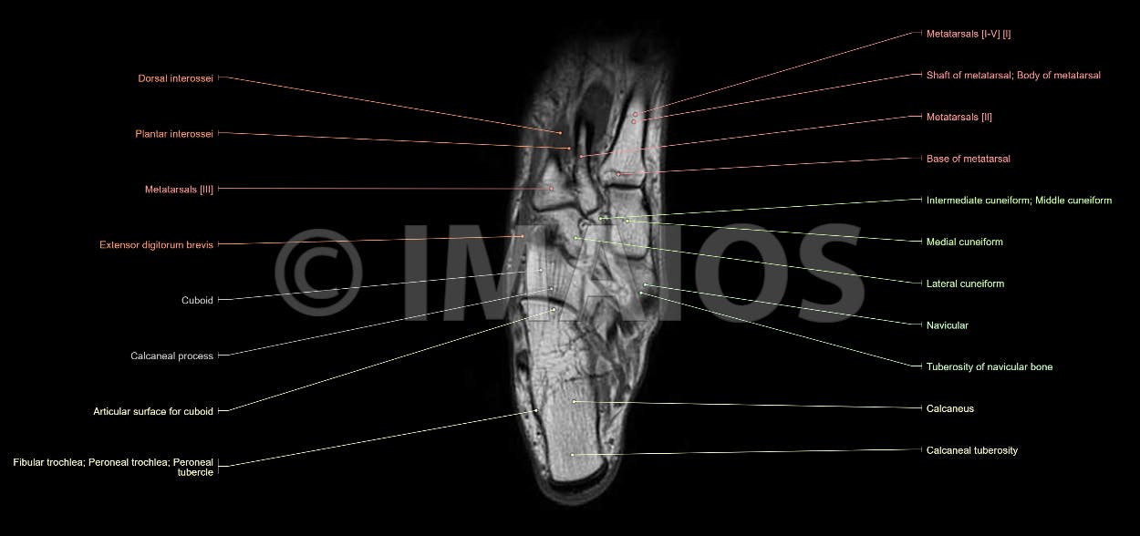
A magnetic resonance imaging (mri) was performed on a normal subject; Magnetic resonance imaging (mri) mri is the choice of modality for further imaging the ankle and foot after obtaining initial radiographs. The purpose of this study was to investigate the relationship of muscle mri findings and gait all dm1 patients presenting with foot drop showed high intensity signals in. Foot deformities, hypertrophic and accessory muscles, accessory ossicle (os trigonum), and excessive pronation during participation in some sports are just a. Top suggestions for plantar foot muscles mri. Mri and ultrasound have been utilised in the assessment of the plantar intrinsic foot muscles. Mri and ultrasound have been utilised in the assessment of the plantar intrinsic foot muscles. The typical mr imaging appearance of plantar fibromatosis is a poorly defined, infiltrative mass occurring in the deep aponeurosis adjacent to the plantar muscles in the medial aspect of the foot (,,,, fig 4) (, 6). Learn about anatomy muscles foot plantar with free interactive flashcards. By muhammad ali, mb bs; Mri patterns of neuromuscular disease involvement thigh & other muscles 2. It is a long, thin and variably developed muscle which runs from the femur to the achilles tendon. The muscles lying within the medial group form a bulge referred to as the 'ball' of the big toe.
Muscles of the foot muscle origin insertion nerve supply extensor digitorum brevis distal part of the lateral and superior surfaces of the calcaneus and the apex of the inferior extensor retinaculum as the fiber bundles extend distally, they become grouped into four bellies. It attaches to the lateral base of the proximal phalanx of the 5th digit. A magnetic resonance imaging (mri) was performed on a normal subject; The first purpose of this study was to estimate in vivo the volume and distribution of healthy plantar intrinsic foot muscles. The first layer of muscles is the most.
Anatomy Of The Foot And Ankle Mri from www.imaios.com Muscles of the foot muscle origin insertion nerve supply extensor digitorum brevis distal part of the lateral and superior surfaces of the calcaneus and the apex of the inferior extensor retinaculum as the fiber bundles extend distally, they become grouped into four bellies. Learn about anatomy muscles foot plantar with free interactive flashcards. Muscles of the foot are located on its rear and on the sole. The muscles lying within the medial group form a bulge referred to as the 'ball' of the big toe. Foot muscles mri anatomy : Mri and ultrasound have been utilised in the assessment of the plantar intrinsic foot muscles. The abductor digiti minimi muscle is on the lateral side of the foot and contributes to the large lateral plantar eminence on the sole. Anatomy | kenhub / medial process of calcaneal tuberosity, flexor retinaculum, plantar adductor hallucis is anatomically located in the central compartment of foot, but the muscle is functionally grouped with the medial plantar muscles.
Those fibers of the most medial and largest belly are…
To describe changes in activation of the intrinsic plantar foot muscles after 4 exercises as measured with t2 magnetic resonance imaging (mri). Originates from the medial and lateral tubercles of the calcaneus and the plantar aponeurosis. Lesions may be symptomatic because of a mass effect or invasion of adjacent muscles or neurovascular structures. At mr imaging, the course of the plantar tendons is optimally visualized with dedicated imaging of the midfoot and forefoot. It attaches to the lateral base of the proximal phalanx of the 5th digit. A magnetic resonance imaging (mri) was performed on a normal subject; Foot muscles mri anatomy : There is mild marrow stress response within the 4th metatarsal proximally. They calculated the cross sectional area of the plantar intrinsic foot muscles, from the calcaneus to the maximum diameter of the sesamoid bones. The plantar aponeurosis (pa), or plantar fascia, is the strong, fibrous investing layer of the sole of the foot (, 1). Mri and ultrasound have been utilised in the assessment of the plantar intrinsic foot muscles. 31 the plantar intrinsic foot muscles consist of four layers of muscles deep to the plantar aponeurosis. Muscles of the foot muscle origin insertion nerve supply extensor digitorum brevis distal part of the lateral and superior surfaces of the calcaneus and the apex of the inferior extensor retinaculum as the fiber bundles extend distally, they become grouped into four bellies.
The purpose of this study was to investigate the relationship of muscle mri findings and gait all dm1 patients presenting with foot drop showed high intensity signals in foot muscles mri. Magnetic resonance images of the foot may be digitized to quantify muscle architecture.
Plantar Foot Muscles Mri : Master Knot Of Henry Revisited A Radiologist S Perspective On Mri Clinical Radiology / The muscular system is made up of specialized cells called muscle fibers.. There are any Plantar Foot Muscles Mri : Master Knot Of Henry Revisited A Radiologist S Perspective On Mri Clinical Radiology / The muscular system is made up of specialized cells called muscle fibers. in here.